Learning objectives:
- The basic anatomy of the heart
- The electrical system of the heart
- How an EKG is performed
- The basic shape of an EKG pattern
- Interpreting a rhythm
Introduction
This semester we have been looking at various phenomena that repeat. One of those phenomena is the contraction of the heart. The record of the electrical signal from the heart’s contraction is called an EKG. On an EKG, we can see both the frequency of the heart’s contraction, which we call the heart rate, as well as the amplitude of the signal.
The anatomy of the heart

The electrical system of the heart

The path of the electrical signal is:
- Signal begins in the sinus node which is fould in the right atrium.
- The signal spreads through both atria, causing the atria to contract.
- The signal passes from the atria to the ventricles through the atrioventricular (AV) node.
- The signal follows two bundle branches from the AV node into the right and left ventricles, causing the ventricles to contract.
The sinus node produces a signal spontaneously. It does not need any signal from the brain or any other part of the body. Below is a link to a YouTube video about heart transplants which shows a heart beating on its own outside of the body. (This video shows blood and may be too gross for some people.)
Transporting a beating heart for transplant – TechKnow
How an EKG is performed

The basic shape of an EKG


Interpreting the rhythm of an EKG
There are many rhythms that can be seen on an EKG. We focused on these:
- normal sinus rhythm
- sinus tachycardia
- sinus bradycardia
- atrial paced rhythm
- supraventricular tachycardia
- atrial fibrillation
- ventricular tachycardia
When looking at a rhythm strip, you need to answer these 3 questions:
- Are there P waves?
- What is the heart rate?
- Is the QRS complex narrow or wide?
Determining a sinus rhythm
P waves that all have the same shape and are all followed by a QRS complex indicate that the heart beat has started in the sinus node. This is called a sinus rhythm.
If there are no P waves or the P waves do not all look the same, then the rhythm is not coming from the sinus node. There may be other places in the heart that are initiating the signal.
Determining the heart rate

Each small square along the horizontal axis represents 0.04 seconds.
Groups of 5 small squares are separated by heavier lines which represent 0.2 seconds.
The heart rate is measured in beats per minute (bpm).
An easy way to estimate the heart rate is by determining how many large boxes fit between two R waves. If only 1 large box fits, then the rate is 300 bpm. If 2 large boxes fit, then the rate is 150 bpm. The diagram above shows the numbers that are memorized to be able to quickly estimate the rate.
The normal heart rate is between 60 and 100 bpm.
If the rate is faster than 100, then the rhythm is a tachycardia.
If the rate is slower than 60, then the rhythm is a bradycardia.
Paced rhythms
Sometimes a person’s sinus node is not working properly. Without a properly working sinus node, the patient’s heart rate may be too slow or too irregular.

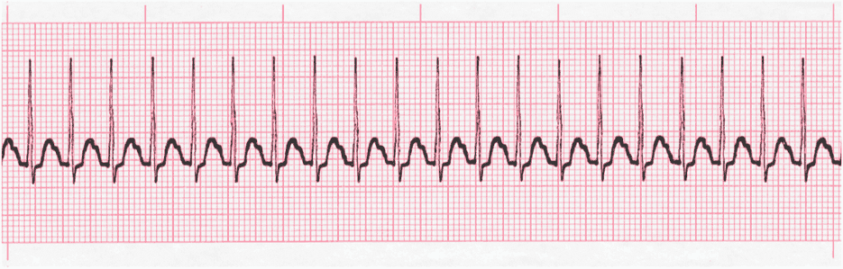
In this image, you can see normal P waves followed by QRS complexes and then T waves. But then there is a gap where another P wave should be, but it is missing. If this happens long enough, it can cause real problems.
When the heart is not beating correctly, an artificial pacemaker can be implanted to take the place of the sinus node.
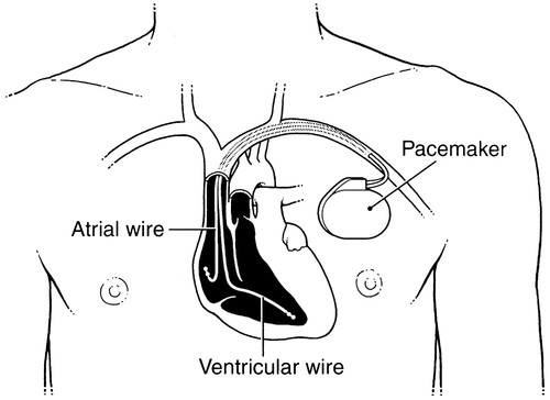
When someone has a pacemaker, there is a sharp spike present on their rhythm strip just before the P wave (or sometimes before the QRS complex).

In this image, you can see the pacemaker spike before each P wave, except the last one which was generated by the sinus node, rather than the pacemaker.
Other Narrow Complex Rhythms
There are other narrow complex rhythms such as:
- SVT
- atrial fibrillation
- atrial flutter
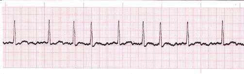
The rhythm above is supraventricular tachycardia.

In SVT, the heart may have an extra pathway that allows the signal to return from the ventricle to the atria, stimulating a new heart beat, faster than the sinus node normally would.
Below is a YouTube video of a patient being treated for SVT with adenosine which blocks the signal at the AV node and breaks the cycle of SVT. The heart stops for a short time and then the sinus node takes over. (This video shows needles and medication being injected into tubing. It may bother some people.)
SVT or Supraventricular Tachycardia
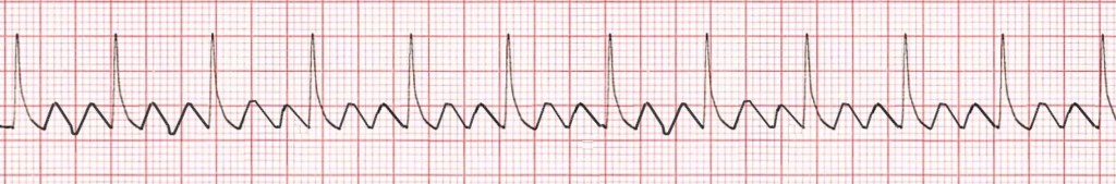
The rhythm above is atrial fibrillation. Atrial fibrillation is important because it is a cause of strokes and heart failure.
The rhythm above is atrial flutter.
Wide Complex Rhythms
When the QRS complex is wide, it often indicates that the rhythm was initiated in the ventricles rather than in the atria.
One such rhythm is ventricular tachycardia.


Leave a comment