- Light Microscopy
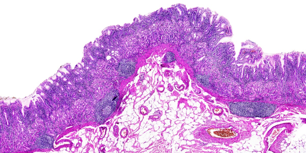
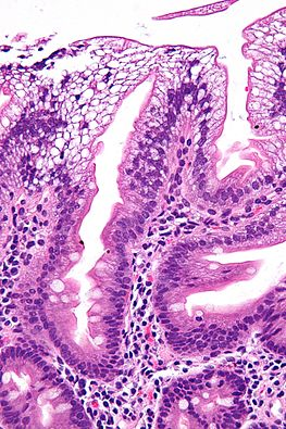
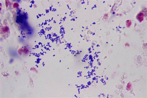
- Staining Techniques
- Stereo (or Dissecting) Microscopy
- Dark Field Microscopy
- Polarized Light
- Phase Contrast
- Fluorescent Techniques
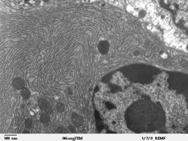
- Transmission Electron Microscopy
- Scanning Electron Microscopy
This post focuses on recognizing what techniques are used to produce certain images. The goal is to help you know what technique was used to create an image that you are looking at.
Light Microscopy
Light microscopy is the most common way to examine microscopic structures. However, a major limitation of basic light microscopy is the lack of contrast. A few structures have natural pigments such as the green chlorophyll in plants and the red hemoglobin in red blood cells. But many cells do not have any pigments. The structures within the cell can be difficult to see.


Staining Techniques
To overcome the lack of natural pigments, specimens are often prepared with a stain. There are many different stains which are used to distinguish different parts of a cell. Staining is commonly used in tissue samples.
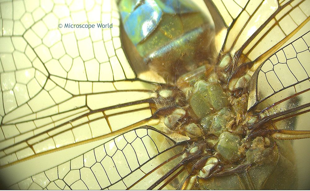
Stereo (or Dissecting) Microscopy
A stereo microscope is often used to look at three dimensional objects.
Dark Field Microscopy
Dark field microscopy is a technique that eliminates background lighting so the objects of interest stand out. These images are distinguished by their dark background.


Polarized Light
Some specimens, especially crystals, change the orientation of polarized light passing through them. This change shows up as different colors and is often useful in determining what type of crystal is being examined. These images are easily distinguished by the swirl of multiple colors.


Phase Contrast
Phase contrast microscopy is another technique used to improve the contrast of an image. These images are distinguished by the halo of light around objects.

Fluorescent Techniques
A specimen can be stained with a fluorescent molecule that binds to a specific molecule in a cell. This allows detailed images of the locations of those molecules. These images are distinguished by their bright colors, which are usually limited to some combination of green, blue and red.


Transmission Electron Microscopy
Transmission electron microscopy (TEM) uses an electron beam, rather than light, to create a 2-dimensional image of a specimen. This provides much higher resolution than light microscopy. Electron micrographs are generated on a computer screen. They are naturally gray-scale images unless they are artificially colored later.
Scanning Electron Microscopy
Scanning electron microscopy (SEM) also uses an electron beam to create a gray-scale image. However, unlike TEM, the electrons are scattered off the surface of the specimen instead of being transmitted through the specimen. Therefore, only the surface of the specimen is imaged. It produces a 3-dimensional image which never has color unless it is a false-color image produced later.


Leave a comment