Upcoming Events and Goals
There is a health science research day on November 15 at the medical school. This will have a lot of students presenting their research. This is done in a room full of students standing next to posters displaying their research and answering questions about their research. There will be a wide variety of research, some of which is fairly straightforward to understand and some of which is overwhelmingly difficult. Fortunately, we are free to choose who we want to interact with. To give you an idea of the range of topics, here is a list of the posters from last year.
I want to help prepare you to be able to interpret their posters so that you can understand the discussion and ask good questions.
Topics we have covered related to this:
- the organization of a research paper (which is also reflected in a poster)
- root words that are used frequently in research
- structural diagrams of organic molecules
- scientific notation
- error bars
- cell membrane receptors
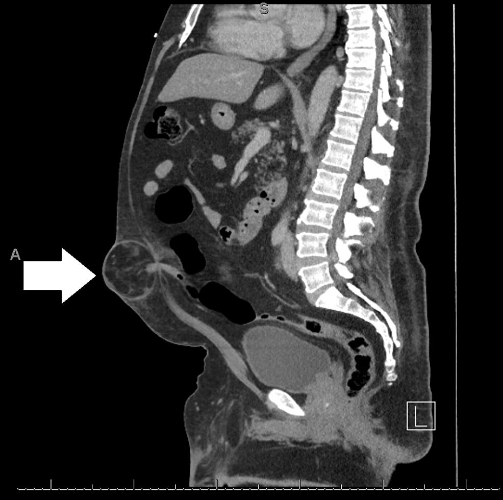
- CT and MRI images
Topics we will need to cover in the next few weeks:
- microscopy pictures
- gel electrophoresis
- genetics
- polymerase chain reaction (PCR)
- model organisms
- reading graphs and charts
- bar charts
- pie charts
- line graphs
- histograms
- semi-log graphs
- log graphs
- Kaplan-Meier plots
- units
- statistics
- p values
- standard deviation
- bell curves
- confidence intervals


Greek and Latin Roots
Here is the list of Greek and Latin roots for this week:
Here is a link to all of the Greek and Latin roots we have discussed.
Test your memory of the Greek and Latin roots that we have discussed with this quiz.
This is the link to the Wikipedia list of Greek and Latin roots.
Presentations
Tips regarding presentations:
- Try to make your topic more specific
- You have the option of doing other things besides an oral presentation.
- You could do a written project
- You could even write an article for this blog
Topics to consider
- How different types of microscopy work, including:
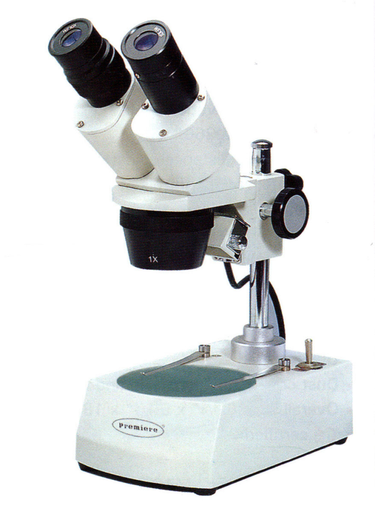
- Stereo microscope
- Bright field
- Dark field
- Phase contrast microscope
- Polarized light microscope
- Confocal microscope
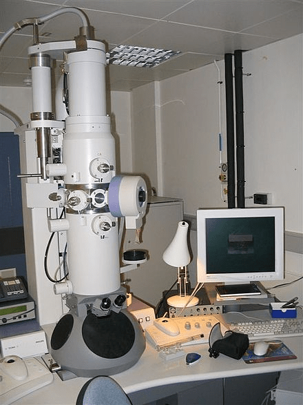
- Electron microscope
- Basic science related to microscopes, including:
- What polarized light is
- Why electrons have a wavelength
- Techniques used in microscopy, such as:
- Gram staining
- Fluorescent antibodies
- The types of herpes viruses and the diseases they cause in people
Assignments
- Fill out the list of Greek and Latin roots.
- Write in the meaning of each root
- Give at least one example of each, be prepared to give its actual definition and the way that it is related to the root word
- Example: If I gave you the root “onym”, you could give the word “synonym” which has the definition of two words with the same meaning. The two roots in the word “syn” and “onym” mean “same name”, indicating two words that name the same thing.
- Fill out the blank space at the bottom with your own root that you have discovered. This will likely come from some of the example words that you have already written. Give a different example than what you have used.
- Example: syn- means “same”, example word “synchronous”
- Be prepared to talk about where you found this information
- Presentation
- Research your topic of choice and be prepared to give a 5 minute presentation on the topic, geared toward people your age level.
- Include the background information needed for someone who does not know the topic as well as you.
- Be prepared to talk about how you found this information.
- Review the page about microscopy images.
Our next meeting will be 10/17/24.
There is a health science research day coming up on November 15.
Things We Discussed
Corrections
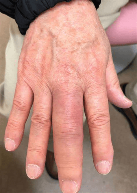
I mentioned that all insect orders use pter-, but this is not true. There are 29 orders of insects and 20 of them include pter-. This is a lot, but not all.
A Guide to the 29 Insect Orders (thoughtco.com)
Greek and Latin roots
Auri – ear
auricle – in addition to the outer ear, the heart also has auricles

Chir – hand, forearm
chiroptera – “hand wing”, the order of bats
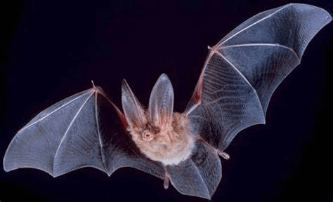
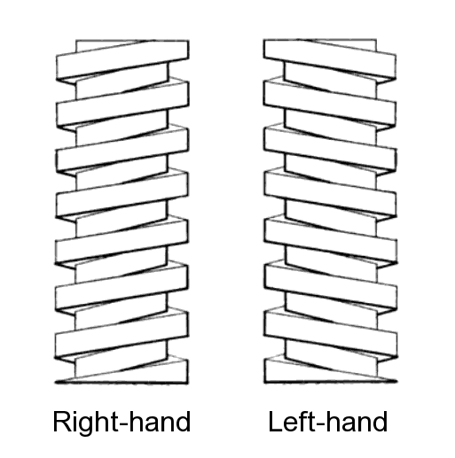
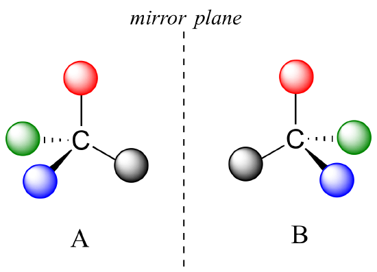
chiral

Cyan- blue
cyanosis

cyanide

Dactyl – finger, toe
pterodactyl – “wing finger”
syndactyly – “digits together”

dactylitis
Labi – lip
nasolabial fold

herpes labialis – “cold sore” caused by a herpes virus

Myo – muscle
myocyte – muscle cell

Pter – wing
Apteryx – “without wings”, genus of bird known as a kiwi

diptera – “two wings” (most insects have 4 wings)
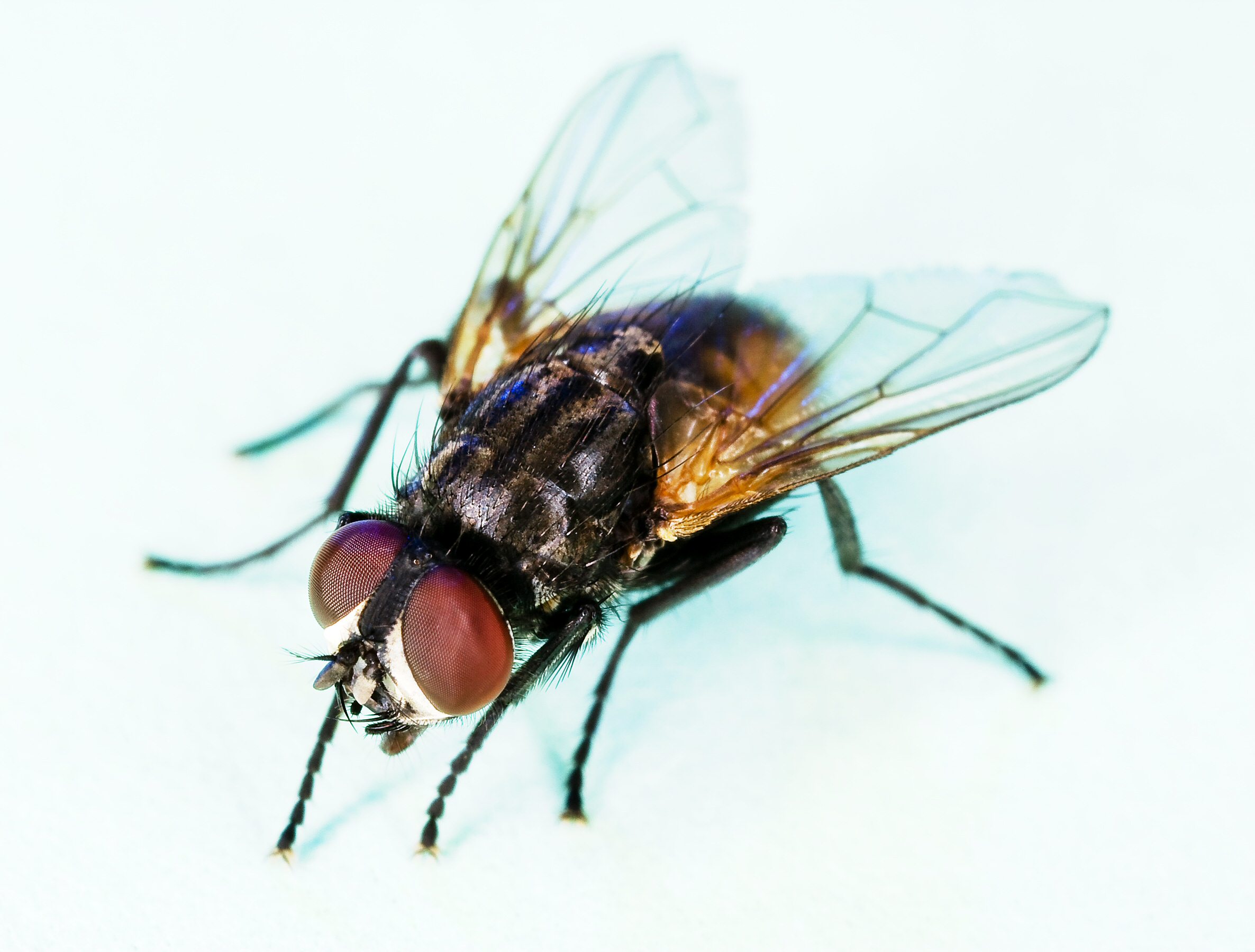
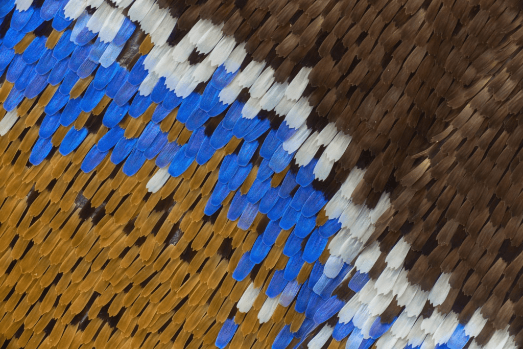
lepidoptera – “scale wing”
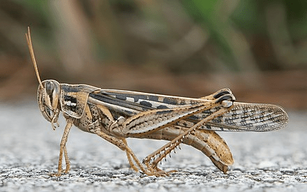
orthoptera – “straight wing”
ornithopter – means “bird wing”, the sci-fi helicopter substitute from the movie “Dune”.

Trich – hair
trichome

achromotrichia – “without color hair”

Umbilic – belly button
umbilical cord

umbilicated

umbilical hernia

Ungu – fingernail, hoof
subungual hematoma
Other roots that were mentioned
- path – disease (mentioned with cardiomyopathy and pathogen)
- a – not, no (mentioned with apteryx)
- chrom – color (mentioned with achromotrichia)
- herp – to creep (mentioned with herpes and herpetology)
Microscopy
Stereo (or dissecting) microscope
Advantages
- Provides a 3-dimensional view of the object being examined
- Able to view bulky objects
- Can work on objects (such as dissecting or assembling) while examining them
Disadvantages
- Magnification limited to 40x
Compound microscope
Advantages
- Capable of high magnification (1000x)
- Can be combined with many other techniques to improve visualization
Disadvantages
- Specimens being examined must be very thin (basically 2-dimensional)

Some techniques that can be used to improve visualization include:
- Staining the specimen
- Changing the way the light interacts with the specimen
- Polarized light
- Dark field
- Phase contrast
Electron microscopes
The resolution of an optical microscope is limited by the wavelength of light. This can be overcome to some extent by using higher frequencies of light, including ultraviolet light or xrays. However, these higher frequencies cannot be directly seen.
Another approach is to use electrons, which also have a wavelength which is much shorter than the wavelengths of light. While electrons cannot be seen, they can be detected to produce a very high resolution image.
Advantages
- Very high resolution (2000 times better than an optical microscope)
Disadvantages
- The sample must be in a vacuum, so wet specimens and living specimens can’t be studied
- Images do not have any color
Micrographs
Pictures taken with a microscope are called micrographs. Micrographs taken with different techniques have different characteristics which allow you to be able to determine what technique was used. It is a useful skill to be able to recognize what technique was used to produce an image.
This page shows examples of different types of micrographs produced using different techniques.
Once you’ve looked over this page, you can take a quiz to test your ability to recognize the different techniques.

Leave a comment