- Upcoming Events and Goals
- Greek and Latin Roots
- Assignments
- Things We Discussed
- Footnotes
Upcoming Events and Goals
There is a health science research day on November 15 at the medical school. This will have a lot of students presenting their research. This is done in a room full of students standing next to posters displaying their research and answering questions about their research. There will be a wide variety of research, some of which is fairly straightforward to understand and some of which is overwhelmingly difficult. Fortunately, we are free to choose who we want to interact with. To give you an idea of the range of topics, here is a list of the posters from last year.
I would like you to consider being there from 8 to 11 am when the undergraduates will be presenting their research. You don’t have to be there the whole time. You will need to talk to your parents about getting out of school and getting there and getting picked up. I’ll be there at 8 to meet you and help you make connections, talk to people and understand their research.
In the meantime, I want to help prepare you to be able to interpret their posters so that you can understand the discussion and ask good questions.
Topics we have covered related to this:
- the organization of a research paper (which is also reflected in a poster)
- root words that are used frequently in research
- structural diagrams of organic molecules
- scientific notation
- error bars
- cell membrane receptors
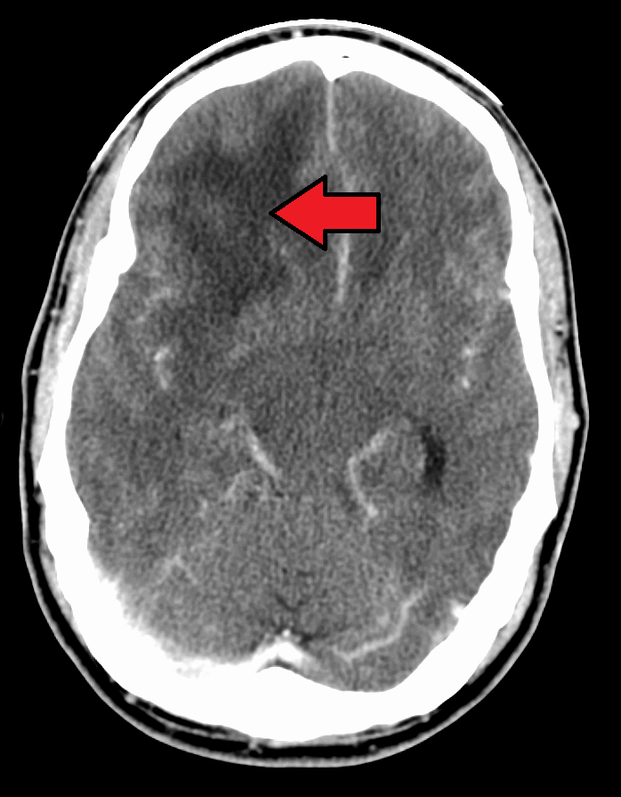
- medical imaging
- antibodies
- microscopy pictures
Topics we will need to cover in the next few weeks:
- gel electrophoresis
- genetics
- polymerase chain reaction (PCR)
- model organisms
- reading graphs and charts
- bar charts
- pie charts
- line graphs
- histograms
- semi-log graphs
- log graphs
- Kaplan-Meier plots
- units
- statistics
- p values
- standard deviation
- bell curves
- confidence intervals
Greek and Latin Roots
Here is the list of Greek and Latin roots for this week:
Here is a link to all of the Greek and Latin roots we have discussed.
Test your memory of the Greek and Latin roots that we have discussed with this quiz.
This is the link to the Wikipedia list of Greek and Latin roots.
Assignments
- Fill out the list of Greek and Latin roots.
- Write in the meaning of each root
- Give at least one example of each, be prepared to give its actual definition and the way that it is related to the root word
- Example: If I gave you the root “onym”, you could give the word “synonym” which has the definition of two words with the same meaning. The two roots in the word “syn” and “onym” mean “same name”, indicating two words that name the same thing.
- Fill out the blank space at the bottom with your own root that you have discovered. This will likely come from some of the example words that you have already written. Give a different example than what you have used.
- Example: syn- means “same”, example word “synchronous”
- Presentation
- Research your topic of choice and be prepared to give a 5-minute presentation on the topic, geared toward people your age level.
- Include the background information needed for someone who does not know the topic as well as you.
- Be prepared to talk about how you found this information.
Next week we will meet on 10/24/24.
Things We Discussed
Greek and Latin roots
ben- good
benign – a meningioma is usually a benign tumor. Benign tumors do not invade surrounding tissue or spread to other parts of the body

mal- bad
malignancy – a malignancy invades into the surrounding tissue or spreads to other parts of the body

a-, an- means no or not
anemia

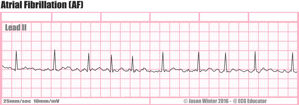
arrhythmia – abnormal heart rhythm4

belli- war
antebellum – before the war (typically the Civil War)

Protein denaturation
Proteins have 4 levels of structure: primary, secondary, tertiary, and quaternary.
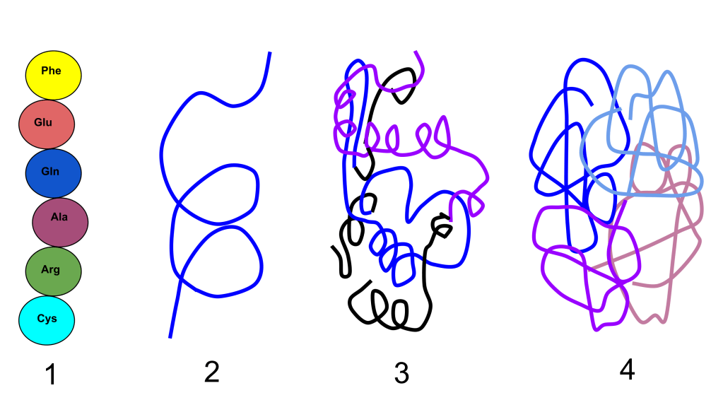
A protein must be folded correctly in order for it to function correctly. Denaturing causes unfolding of the protein leading to loss of function. Denaturing can be caused by heat, by change of pH, or by addition of a chemical. Denaturing can cause loss of the quaternary, tertiary or even secondary structure, but does not break the bonds between the amino acids, so the primary structure remains intact. Sometimes denaturing is reversible and sometimes it is irreversible. The classic example of denatured proteins is the white of an egg when it is cooked.
The secondary structure of the protein is caused by hydrogen bonding between hydrogen atoms and oxygen atoms in the amino acid chains.5

Changing the pH (making the solution more acidic) increases the number of hydrogen ions in the solution. These hydrogen ions can be attracted to the oxygens, breaking up the hydrogen bonds and disrupting the secondary structure of the protein.
Blood flow through the heart

The blood in the left and right sides of the heart does not mix. In fact, the left and right sides can be thought of as two different hearts. The diagram below is a little complicated, but you can see that the main idea is a single loop: body -> right heart -> lungs -> left heart -> body
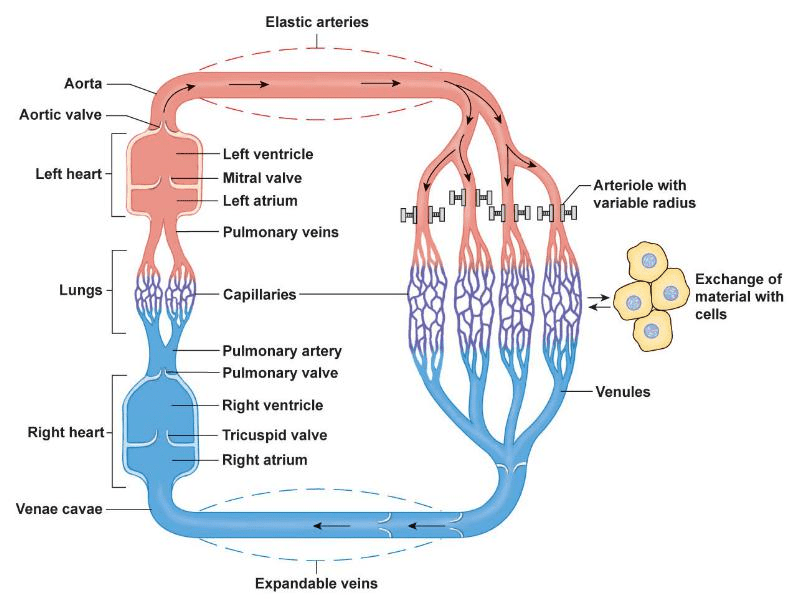
There are two benefits to having both halves of the heart together. One is that the electrical system in the heart sends signals to coordinate both halves to beat at the same time. The other is that the septum does not have to be as strong, because the pressure from the right half squeezing balances the pressure of the left half squeezing.
Antibodies
Blood cell lines
Blood cells are produced in the bone marrow, starting with a stem cell that is able to turn into various “blast” cells. When the blood cells are mature, they move from the bone marrow to the blood stream.

Although this diagram looks more complicated, blood cells can be thought of as having three different types:
| Blood Cell Type | Function |
| Erythrocyte (Red blood cell) | Carries oxygen |
| Leukocyte (White blood cell) | Fights infection |
| Thrombocyte (Platelet) | Forms blood clots |
White blood cells are classified as either myeloid cells or lymphocytes.
Lymphocytes
There are three types of lymphocytes: B cells, T cells and natural killer cells. The ones we are interested in are B cells. B cells produce antibodies.
The human body contains millions of B cells which all produce a different type of antibody. Before an antigen attaches to the B cells, the B cell only has antibodies on its surface. Once an antigen binds to the B cell, the B cell is activated and converts into a plasma cell, producing large amounts of antibodies that it puts into the blood stream.
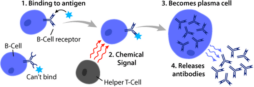
Antigens
An antigen is any molecule that can cause an immune response. Typically, an antigen is a protein or polysaccharide molecule6. Very small molecules cannot be antigens. For instance, you cannot develop an immune response against a water molecule.
Structure of antibodies
An antibody is a protein that is made of 4 strands of amino acids. There are two bigger polypeptides (called heavy chains, yellow and blue in the diagram) and two smaller polypeptides (light chains, green and red in the diagram). These 4 polypeptides bind together to make one protein. The antibody has two binding sites where an antigen can attach.

To simplify drawings, an antibody is typically drawn as a Y-shaped structure with two longer lines and two smaller lines.

Classification of antibodies
There are 5 classes of antibodies in humans:
| Class | Function |
| IgA | found on mucosal surfaces (wet surfaces) like the inside of your nose, mouth, lungs, gut and urinary tract. These fight off infection before it actually invades. |
| IgD | these act as B cell receptors on B cells before they become activated and form plasma cells |
| IgE | cause allergic reactions, but also helps fight parasitic infections |
| IgG | this is the main class of antibodies that fight off infection in the blood stream |
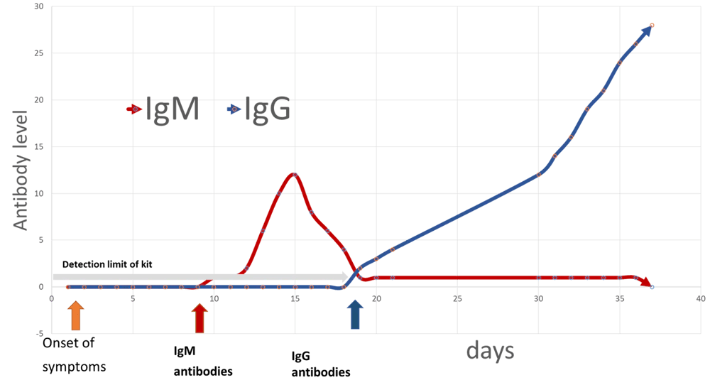
| IgM | these are formed immediately after infection and then drop off as IgG becomes the dominant antibody later |
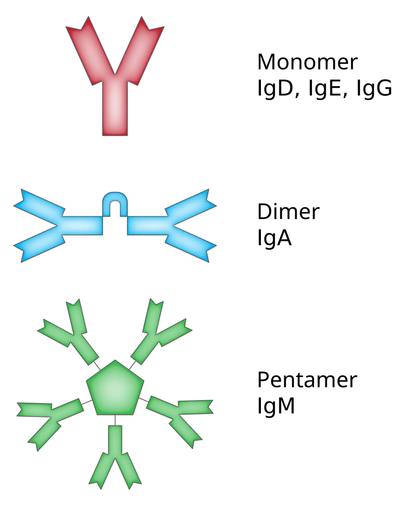
Antibodies may stick together to form dimers (IgA) or pentamers (IgM).
Passive vs. active immunity
If a person is exposed to an antigen and they begin producing antibodies against the antigen, this is called active immunity.
If instead, a person is given an antibody, this is called passive immunity. These antibodies will eventually break down and the person is not able to make more of those antibodies because they were never exposed to the antigen.
Examples of passive immunity:
- IgG passes through the placenta from the mother to the baby to give the baby an immune system when it is first born
- IgA is passed from the mother through breast milk to the baby
- Immunoglobulin against rabies is given along with a rabies vaccine to protect a patient until they are able to develop their own antibodies to the vaccine
- Rattlesnake antivenom is produced by injecting rattlesnake venom into a horse which then produces antibodies that are injected into a snakebite victim to neutralize the venom.
Examples of active immunity:
- A person gets a viral infection and then develops antibodies against the virus so they will not be susceptible to the infection again
- A person is given a vaccination against a disease and develops antibodies against the antigens in the vaccine
Autoimmune diseases
Usually, your immune system is able to distinguish between antigens that are part of your body (“self”) and antigens that come from outside your body (“foreign”). This ability of distinguishing “self” from “foreign” is very important to keep your immune system from attacking your own body. Unfortunately, sometimes things go wrong, and a person can develop an autoimmune disease, in which they develop antibodies that attack their own cells.
One example of this is type I diabetes mellitus7. The level of glucose (blood sugar) in the blood is controlled by insulin which is produced by specialized cells in your pancreas called beta cells. If all of the beta cells are destroyed, then a person cannot produce insulin, and they develop type I diabetes. One way that these cells can be destroyed is by developing antibodies against the beta cells.8
Monoclonal antibodies
Monoclonal antibodies are antibodies that are all produced by one type of cell that is created in the lab for a specific use.
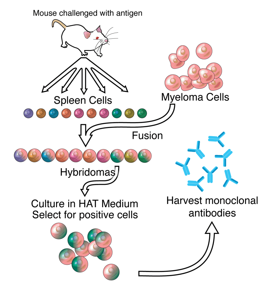
Steps to producing monoclonal antibodies:
- A mouse or rabbit is injected with an antigen which stimulates it to develop plasma cells that produce antibodies against that antigen.
- The plasma cells are removed from the animal.
- The plasma cells are merged with cancer cells to produce hybridomas, which continue to produce antibodies, but now survive and reproduce in the laboratory like cancer cells. These many different cells are making many different antibodies that all bind to the same antigen. These antibodies would be “polyclonal”.
- A specific cell that makes antibodies against the antigen is selected and cloned into millions of cells. All of these cells will produce identical antibodies. Because these antibodies are produced by clones of a single cell, they are called “monoclonal”.
There are currently over 500 different monoclonal antibodies that have been developed.9
Conjugation
The antibodies are often linked to another molecule. This linking process is called conjugation.
Antibodies are often conjugated to:
- a fluorescent molecule
- an enzyme that causes a color change due to a chemical reaction
- a molecule containing a radioactive atom
The molecules that are conjugated to the antibodies are often used to be able to visually detect the presence of the antibodies.
Uses for antibodies
Because of the unique ability for antibodies to be very specific about what they attach to, and to be made to attach to almost anything, they have many uses in laboratory science and medical therapy.
Some of these uses include:
- Testing
- Purification
- Treatment
ELISA
ELISA stands for enzyme-linked immunosorbant assay.

This test allows for rapid testing of many samples at one.
Rapid Antigen Tests


Common rapid antigen tests include:
- Covid infection
- Strep throat
- Pregnancy
Immunofluorescence
Immunofluorescence is a microscopy technique that allows specific structures within a cell to be visualized by binding antibodies that are conjugated to a fluorescent molecule. After the antibodies attach to the structure in the cell, the structure can be visualized by shining ultraviolet light on it, causing the fluorescent molecule to give off light that can be seen under a microscope.

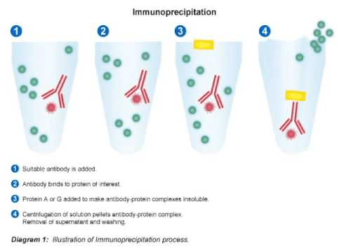
Purification
One method of using antibodies to isolate and purify a sample is called immunoprecipitation.
Monoclonal antibody therapy
Monoclonal antibodies can be used as medical therapy in many diseases including:
- Infection
- Autoimmune disease
- Cancer
Drug names of monoclonal antibodies have an invented root “-mab” at the end of their name to indicate they are monoclonal antibodies. For instance, infliximab is a monoclonal antibody that targets an inflammatory molecule called TNF-α10 and is used to treat autoimmune diseases such as rheumatoid arthritis, ulcerative colitis, Crohn disease and ankylosing spondylitis.
Footnotes
- Medical imaging (Basic science review) ↩︎
- Medical imaging (Basic science review) ↩︎
- Medical imaging (Basic science review) ↩︎
- EKG ↩︎
- Molecular diagrams ↩︎
- Polymers ↩︎
- Type II diabetes mellitus is due to insulin resistance. Your pancreas makes enough insulin but the cells that should respond to the insulin don’t response as well as they should. There is another type of diabetes, diabetes insipidus, which does not have anything to do with blood sugar levels. (“mellitus” refers to the urine tasting sweet due to the glucose in it; “insipidus” refers to the urine not tasting sweet. Fortunately, we have better tests now than tasting urine.) ↩︎
- Other ways to develop type I diabetes include having surgery to remove a pancreatic cancer, developing chronic pancreatitis (from drinking too much alcohol, for instance), some chemicals and chemotherapy treatments, and cystic fibrosis. ↩︎
- See this page for a full list: List of therapeutic monoclonal antibodies – Wikipedia ↩︎
- TNF stands for “tumor necrosis factor” ↩︎

Leave a comment