Radiology is the use of various imaging techniques to understand what is going on in the body. There are many different imaging techniques that are used, including:
X-ray
- An x-ray is an image obtained by exposing a screen to x-rays that have passed through the body. Some of the x-rays are absorbed by the body. Denser regions of the body are more white and less dense regions are darker.
- X rays are ionizing radiation that can cause cancer.

CT scans
- CT stands for computer-aided tomography. So, sometimes they are called CAT scans.
- CT scans take a lot of pictures using x-rays from multiple angles. This exposes the patient to a lot of ionizing radiation1 which can cause cancer.
- It also provides a very detailed picture of the insides of a patient.

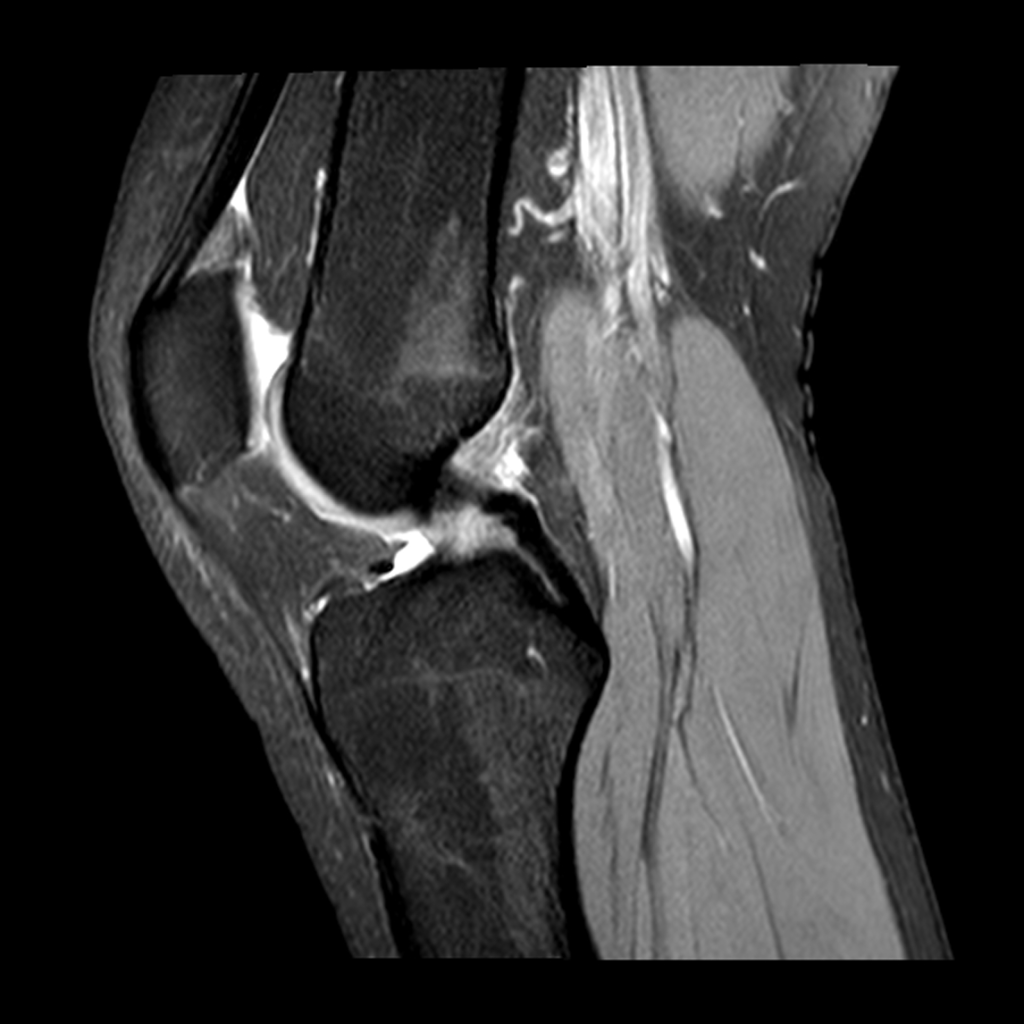
MRI
- MRI stands for magnetic resonance imaging
- MRI does not use ionizing radiation and so will not cause cancer
- The process of obtaining an MRI is significantly slower than getting a CT scan
PET scans
- PET stands for positron emission tomography
- Positrons are antimatter particles that have the same mass as an electron but opposite (positive) charge
- When a particle and an antiparticle collide, they destroy each other and turn into gamma rays2.
- To prepare for a PET scan, a patient is injected with a molecule that has a radioactive atom in it.
- The molecule is absorbed by cells and are concentrated in cells that require a lot of energy, such as cancer cells, but also the brain and heart. It is also excreted in the urine and so the kidneys and bladder normally are highlighted.
- When the radioactive atom decays, it emits a positron. The positron soon hits an electron. The positron and electron destroy each other and produce 2 gamma rays. The gamma rays travel in opposite directions.
- The PET scanner detects the gamma rays and can calculate where they came from in the body by time difference of when they are detected.

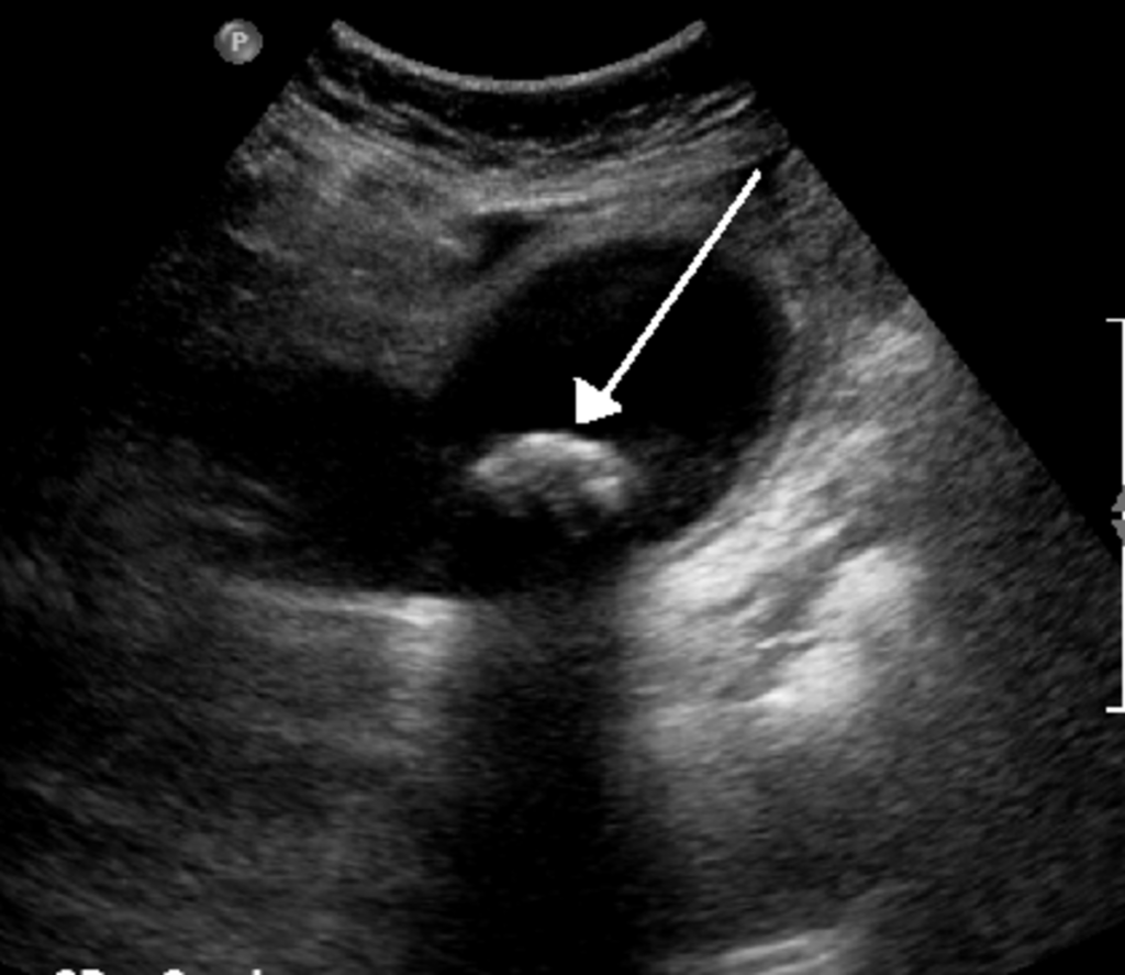
Ultrasound
- An ultrasound image is produced by recording ultrasonic sound waves that bounce off the tissues in the body.
- Ultrasound is particularly good at imaging certain organs, including the gall bladder and ovaries.
- Ultrasound is also very good for imaging a fetus because it does not use ionizing radiation.

Leave a comment