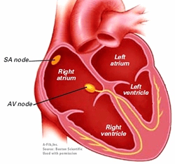
What is this?
This is a cross-section of a heart. It is likely meant to represent a human heart, although at this level of detail it could be any mammalian or avian heart. However, reptilian hearts have only 3 chambers. (Crocodilians have 4 chambers but an extra feature that causes mixing of oxygenated and deoxygenated blood.)
Why are the right atrium and right ventricle shown on the left?
The illustration shows the heart as if you are looking at someone else’s heart. This is standard practice for anatomic diagrams showing the anterior view.
What are the vessels that are shown?
- At the left, the superior vena cava enters the right atrium from the top and the inferior vena cava enters from the bottom.
- On the far right, the pulmonary artery carries blood from the right ventricle to the lungs.
- Between the superior vena cava and the pulmonary artery is the aorta which carries blood from the left ventricle to the body.
What is the SA node?
The sinoatrial node is the location where the natural pacemaker cells of the heart send out the signal for the heart to beat.
What is the AV node?
The atrioventricular node conducts the electrical signal from the atria to the ventricles.
What are the yellow lines coming from the AV node?
These are the fibers (known as the left and right bundles) which carry the electrical signal from the AV node to the left and right ventricles.
Why is the left ventricle smaller than the right ventricle?
The volume of blood that passes through the right and left ventricles with each heartbeat must be the same. However, the right ventricle does not empty as completely as the left ventricle. Therefore, the volume of the right ventricle is slightly greater than the left ventricle.
But the main reason the illustration shows the right ventricle is larger is because the heart is a 3-dimensional structure and the cross-section through the heart that is being shown just goes more directly through the right ventricle than it does through the left.
What other structures should you be able to identify in this illustration?
Other structures you should identify in this illustration include:
- The mitral valve between the left atrium and ventricle.
- The tricuspid valve betwen the right atrium and ventricle.
- The atrial septum separating the two atria.
- The ventricular septum separating the two ventricles.
- The relative thickness of the walls of the atria (which are thin), the right ventricle (which is thicker) and the left ventricle (which is the thickest).

Leave a comment