Where are these structures located in the body?
The kidneys are in the back of the abdomen at about the level of the lowest ribs.
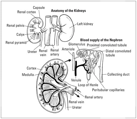
What distinguishes the cortex and medulla?
The cortex is the outer layer and the medulla is the inner layer. There are several organs that are organized this way including the adrenal glands, thymus, ovaries, lymph nodes, cerebrum, cerebellum, and spinal cord. In each of the organs, the cortex serves a different function and is organized a different way than the medulla.
In the case of the kidney, the cortex holds the glomeruli and the medulla contains the loops of Henle and the collecting ducts. The arterioles and venules are at the boundary between the cortex and medulla.
What is mislabeled in this image?
In the top picture, the structures labeled “renal vein” and “renal artery” are actually the inferior vena cava and the aorta. The renal artery and vein are correctly labeled in the bottom picture.
What explains the asymmetries between the kidneys?
The right kidney is slightly lower than the left because the liver is on the right side of the body.
The left renal vein is longer than the right because it has to cross the aorta to get to the inferior vena cava.
How could this asymmetry affect the blood supply of the kidneys?
The renal vein can sometimes become pinched between the aorta and the superior mesenteric artery, restricting blood flow from the left kidney.
What is drawn incorrectly in this image?
The right renal artery is not shown in the top image. Instead, there are two vessels running from the right kidney to the inferior vena cava. A duplication of the right renal vein can actually happen. It is important to see that those two vessels drawn in the image do not actually represent the renal artery and vein, but rather two veins.
At what level does the arterial and venous systems stop being parallel to each other?
The arteries and veins run parallel to each other until the arteriole and venule level where the arteriole enters the glomerulus and the
As a general rule, arteries and veins run parallel to each other in each organ although there are often variations. One important exception is the blood supply of the intestines. The mesenteric arteries come directly from the aorta; however, the mesenteric veins connect to the portal vein which takes the blood from the intestines to the liver.
What is the 3-dimensional structure of the kidney?
The renal pyramids are all pointing inward toward the pelvis. They are cone shaped, rather than pyramidal with flat sides.
The collecting tubules will have nephrons emptying into them from all directions.
The blood vessels branch into a 3-dimensional structure at the renal pelvis, rather than when they reach the cortex.
What are the fluids involved and where are they?
Blood is in the arteries, veins, arterioles, venules and capillaries. Urine is in the proximal convoluted tubules, loops of Henle, distal convoluted tubules, collecting ducts, calyces, renal pelvis and ureters.
Goals of this image:
- Use what your knowledge of human anatomy to help interpret this image
- Location within the body
- Aorta and inferior vena cava
- Asymmetry due to the liver
- Introduce you to the details of renal anatomy
- Cortex and medulla
- Structure of the internal blood supply
- Structure of the nephron
- Envision the actual structure of the kidney
- Deducing the 3-dimensional structure from a 2-dimensional drawing
- Deducing what tubes are connected
- Point out the risk for errors in anatomical drawings
- Mislabeled aorta and inferior vena cava
- Missing right renal artery

Leave a comment